

Our Vision
To establish the Department of Anatomy as a centre of excellence in anatomical sciences that develops knowledgeable, observant and responsible medical professionals with strong foundations for clinical practice.
Explore human structure through dissection, histology, embryology, imaging and clinically relevant anatomical learning
The Department of Anatomy at Autonomous State Medical College, Lalitpur provides students with a strong foundation in understanding the structure, organization and development of the human body. Anatomy forms an essential part of medical education and prepares students to interpret normal body structures before progressing to clinical subjects and patient-based learning.
The department follows a structured teaching approach that combines classroom lectures, cadaveric dissection, practical demonstrations, histology sessions, museum-based learning and small-group discussions. These learning methods help students understand anatomical relationships while developing observation, interpretation and practical skills. Models, specimens, bones, charts, microscopes and medical images are used to make complex concepts easier to understand.
The academic curriculum covers general anatomy, gross anatomy, histology, embryology, neuroanatomy, genetics, surface anatomy, radiological anatomy and clinically applied anatomy. Students are encouraged to connect theoretical concepts with physical examination, diagnostic imaging, surgical procedures and common clinical conditions. This integrated approach helps them understand the importance of anatomical knowledge in future medical practice.
Faculty members guide students through regular teaching sessions, practical exercises, tutorials and assessments. Continuous feedback helps learners identify areas for improvement and build confidence in both theoretical and practical understanding. The department promotes academic discipline, professional conduct, respect for the human body and responsible learning.
Through systematic instruction and clinically relevant teaching, the Department of Anatomy aims to prepare students with the knowledge, skills and professional values required for their continuing journey in medical education.
 Preclinical Department
Builds the foundation for clinical medical learning.
Preclinical Department
Builds the foundation for clinical medical learning.
 Phase I
Taught during the first phase of MBBS.
Phase I
Taught during the first phase of MBBS.
 Dissection Hall
Supports supervised dissection and practical anatomical study.
Dissection Hall
Supports supervised dissection and practical anatomical study.
 Anatomy Museum
Provides specimens and models for visual learning.
Anatomy Museum
Provides specimens and models for visual learning.
The principles guiding the Department of Anatomy in building strong scientific foundations, practical understanding, ethical conduct and clinically relevant medical knowledge.
To establish the Department of Anatomy as a centre of excellence in anatomical sciences that develops knowledgeable, observant and responsible medical professionals with strong foundations for clinical practice.

To provide competency-based anatomy education through lectures, cadaveric dissection, histology, demonstrations, medical imaging and clinical correlation while encouraging scientific curiosity, practical competence, respectful learning and continuous academic development.

The Anatomy curriculum combines theoretical knowledge, practical exploration and clinical application to help Phase I MBBS students understand the structure and development of the human body.
Covers regional anatomy, cadaveric dissection, surface landmarks and structural relationships across major areas of the human body.
Develops an understanding of microscopic tissues, organ development, congenital variations and their importance in clinical medicine.
Connects anatomical knowledge with clinical examination, radiological images, common injuries, surgical procedures and diagnostic interpretation.
Department of Anatomy combines classroom instruction, practical training, visual resources and continuous assessment to develop strong anatomical understanding and clinical application.
Structured lectures use illustrations, presentations, models and clinical examples to explain important anatomical concepts clearly and systematically.
Supervised dissection sessions help students directly observe anatomical structures, understand their relationships and develop essential practical knowledge.
Microscope-based practical sessions enable students to identify normal tissues, organs and their detailed cellular and structural organization.
Anatomical specimens, models, bones and charts support visual learning, concept revision and a deeper understanding of human structures.
Tutorials, demonstrations and group discussions encourage active participation, problem-solving, collaborative learning and clarification of complex anatomical topics.
Regular tests, viva examinations, practical assessments and constructive faculty feedback help students evaluate progress and improve academic performance.
Meet the faculty and staff supporting anatomy education through classroom teaching, practical guidance, academic mentoring and effective departmental administration.
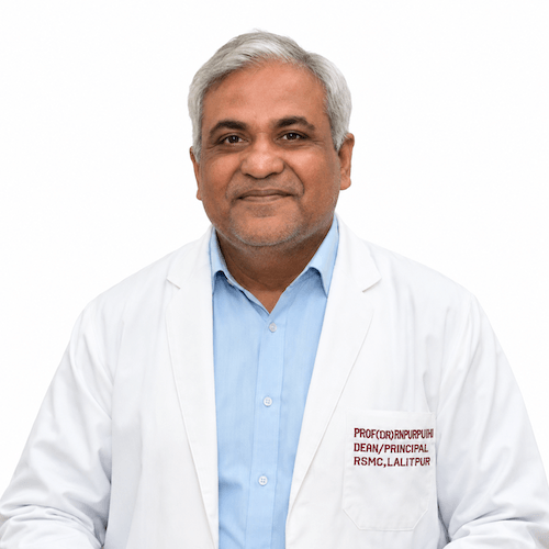
Principal
MBBS, MS (Orthopaedics)

Vice Principal
Qualification awaiting

Chief Medical Superintendent (CMS)
MBBS

Medical Superintendent
Professor, Department of Microbiology
Explore the facilities and learning resources that support observation, demonstrationlk demonstration, practical training and a deeper understanding of human anatomy.

Supports supervised cadaveric dissection and detailed practical study of anatomical structures and relationships.

Provides anatomical specimens, models and charts for visual understanding, demonstration and academic revision.

Supports microscopic examination of normal tissues, organs, cells and their structural organization.

Provides an interactive space for demonstrations, tutorials, discussions and focused small-group teaching.

Supports the systematic study of bones, joints, landmarks and their clinical importance.

Enhances anatomy education through medical images, presentations, illustrations and technology-supported learning materials.
Students are assessed through theory examinations, practical exercises, spotters, viva examinations, internal assessments and regular faculty feedback according to academic regulations.
The curriculum covers general anatomy, gross anatomy, histology, embryology, neuroanatomy, genetics, surface anatomy, radiological anatomy and clinically applied anatomy.
Yes. Supervised cadaveric dissection helps students observe anatomical structures directly and understand their positions, relationships and clinical significance.
Students learn through anatomical specimens, models, bones, charts, microscopes, histology slides, medical images and practical demonstrations.
Anatomy provides the structural foundation required to understand physical examination, medical imaging, injuries, surgical procedures and other clinical subjects.